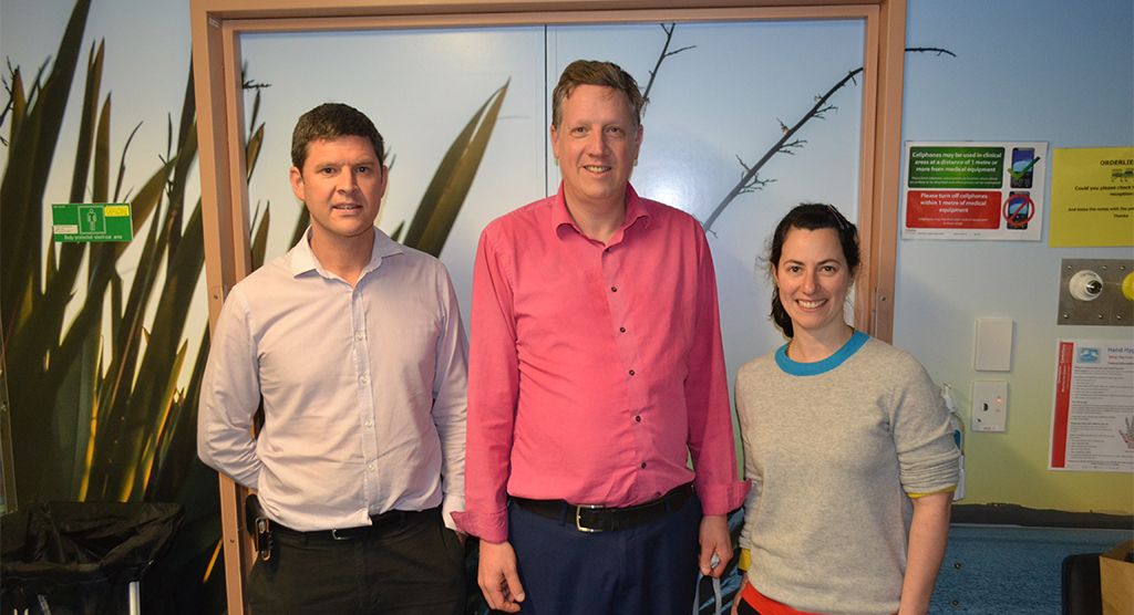
New Zealand clinicians deliver patient-friendly SABR lung in under two minutes
Jul 12, 2023
We want to ensure optimum use of our website for you, and to continually improve our website. Therefore, we work with selected partners (e.g. Pardot, Google Analytics, Matomo). You can revoke your voluntary consent at any time. You can find further information and setting options under "Configure" and in our data protection information.

Are you using all the clinical imaging features that your Elekta XVI® is capable of?
Your XVI system is capable of providing a whole range of advanced imaging tools to help you enhance the visualization of targets and OAR, and ultimately improve clinical accuracy and precision. Reach out today and find out how you can start providing these benefits to patients today.
Enhance your imaging capabilities today with tailored, patient-friendly solutions suitable for the task at hand.
4D kV imaging
Treat moving targets without compromise
Our unique 4D kV imaging solution is called Symmetry. It doesn’t require additional devices—like external surrogate markers and cameras—to create a 4D CBCT. Symmetry uses anatomical correlation, meaning all the 4D information comes from the patient’s own daily anatomy. Simple!
Having Symmetry means that organs moving due to respiratory motion will appear clear. This allows moving targets to be treated aggressively without compromising the safety of adjacent critical structures.

A small SBRT lung lesion near the diaphragm is virtually invisible without 4D image guidance.
St. James Institute of Oncology
Leeds Teaching Hospitals, NHS Trust, Leeds, the UK
We know where the tumor is located every day, for every fraction. We would not perform SABR lung treatments without it.
Dr. Andy Cousins
Christchurch Hospital, New Zealand
Critical structure avoidance
Critical Structure Avoidance, or CSA, allows you to do two image registrations (matches) on one VolumeView image. Its automatic, protocol-driven registration of the target and critical structures helps you minimize dose to OAR while maximizing dose to the target.


Intra-fraction imaging
Why pause your patient’s SABR treatment to acquire a verification CBCT?
With XVI Intra-fraction imaging you can:
3D automated seed matching
Our optimized 3D registration algorithm allows you to register implanted markers, without compromising on 3D volumetric information.
It has been clinically tested to automatically locate:

Dual registration is a unique technique within Elekta CT-Linacs that allows for the registration of images in radiation therapy. It can be used to register a 3D/4D CBCT image to a reference CT. It can also be used to register multiple targets at once.
The key features include:
Clipbox registration
Corrects positioning deviation
Mask registration
Corrects motion deviation
Critical Structure Avoidance (CSA)
Automatically registers the target and critical structures to
minimize dose to OAR while maximizing dose to the target
What are the possible benefits of dual registration?
Protects critical structures
Allows for the protection of critical structures like ribs
Improves treatment accuracy
Allows for the real-time monitoring of tumor movement during treatment
Shortens treatment time
Allows for multiple targets to be treated simultaneously and
effectively
Reduces the risk of positioning errors
Allows for the treatment of multiple targets simultaneously and
effectively

Clinical demonstration
Do you want to improve visualization, benefit from more automation and enable new treatment techniques for your patients? Reach out today and your Elekta representative will reach out to discuss your options.