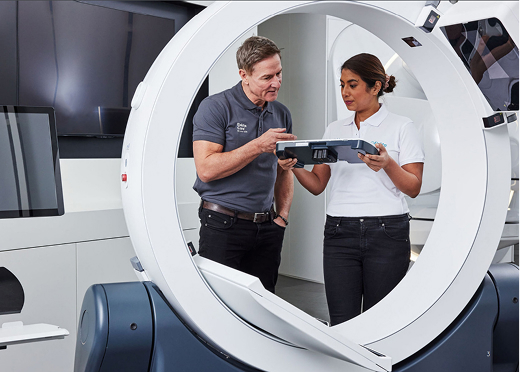
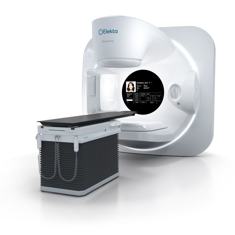
Elekta Harmony
The perfect balance, without compromise
- Unlock perfect productivity
- Elevate precision with versatility
- Experience more than a linac
We want to ensure optimum use of our website for you, and to continually improve our website. Therefore, we work with selected partners (e.g. Pardot, Google Analytics, Matomo). You can revoke your voluntary consent at any time. You can find further information and setting options under "Configure" and in our data protection information.
At Elekta, our journey in in-room imaging began with a promise: to deliver precision that transforms care.
Through our 20-year legacy in imaging excellence, we have continuously advanced technology to empower clinicians to deliver truly personalized treatment.
It’s all about boosting clinician confidence and patient comfort, strengthening trust in the treatment process.
In-room imaging transforms radiotherapy by providing a real-time view of the patient’s anatomy during treatment. This ensures verification of the target, even with changes in the tumor size and shape or surrounding tissue changes.
With our innovations, you can adapt to these changes, delivering accurate radiation while protecting healthy tissue to preserve quality of life.
These patient stories reflect what’s possible when cutting-edge technology and clinical expertise come together.
Innovation alone isn’t enough—it’s the partnership between our solutions and the expertise of clinicians that truly transforms lives.
We’re proud of what we’ve achieved together. Our colleagues across Elekta have shared how imaging makes a real difference for patients

See how our innovations adapted to cancer
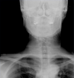
2D imaging
But... no soft tissue visualization
Fluoroscopic-like sequence visualizes moving targets to show organ motion in real-time. Often internal or external markers have been used as a surrogate for tumors, but these can be time consuming and invasive.
3D kV imaging
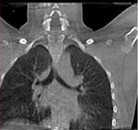
Soft tissue detail but no motion
3D kV imaging enables volumetric visualization to provide soft tissue detail of the target and critical structures in 3D.
Soft tissue information but no motion, this limited information results in uncertainty...
4D kV imaging
Treat moving targets without compromise
Symmetry, what we call our 4D Kv imaging, doesn't require additional devices—like external surrogate markers and cameras—to create a 4D CBCT. Symmetry uses anatomical correlation, meaning all the 4D information comes from the patient's own daily anatomy.
CBCT Gamma Knife imaging
Deliver treatments with unmatched accuracy
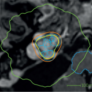
Integrating stereotactic CBCT directly into the Leksell Gamma Knife workflow, enables precise 3D localization to support frameless planning and fractionated treatments. CBCT images taken before treatment serve as the stereotactic reference, with CBCT scans at the time treatment.
MR imaging
Soft tissue detail but no motion
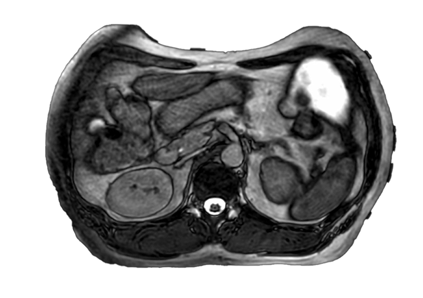
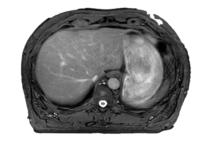
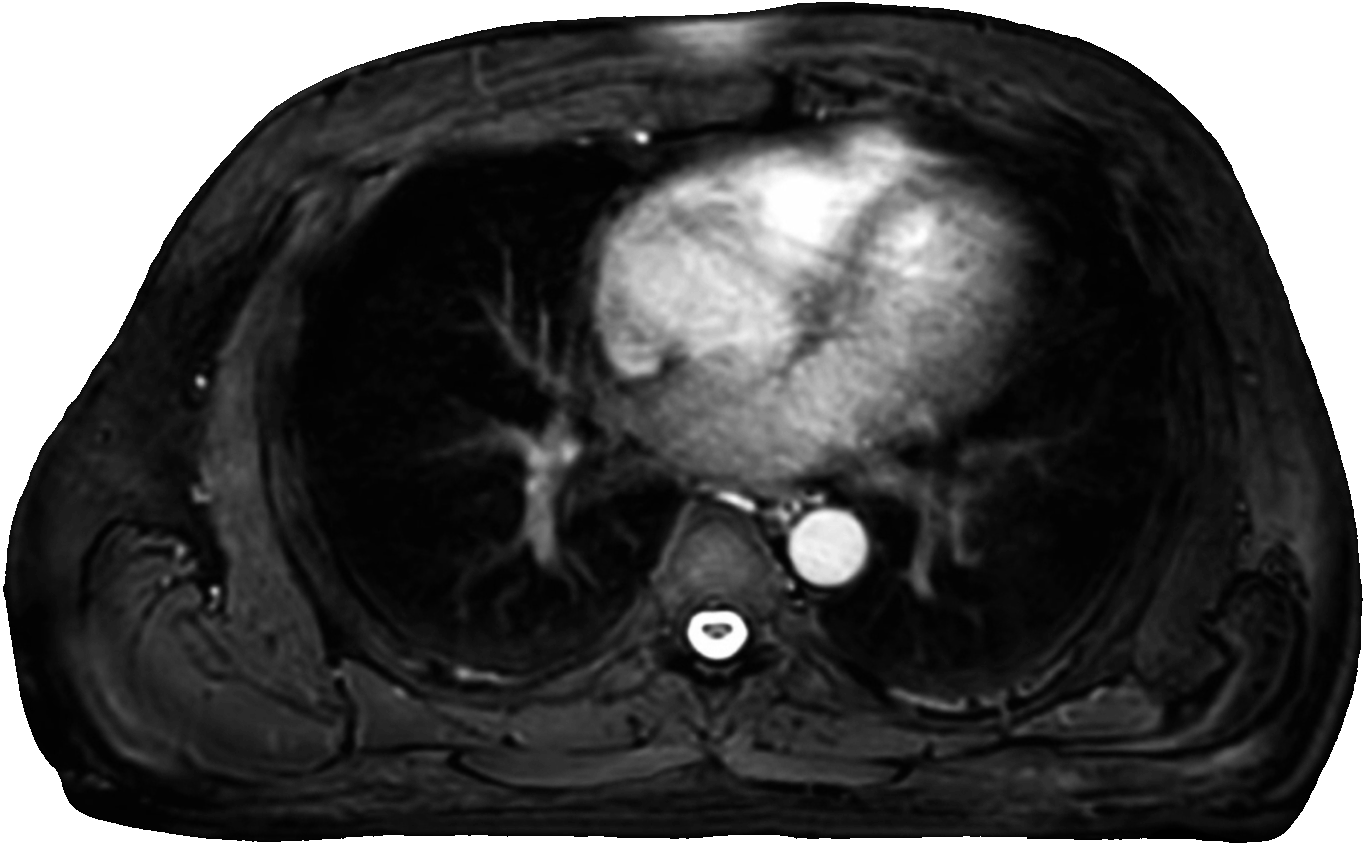
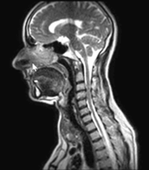
Elekta Unity combines diagnostic quality MRI with precise radiation delivery—a unique combination which provides superior soft tissue visualization of the relationship between the tumor, and surrounding anatomy allowing you to confidently escalate daily dose.
CBCT in brachytherapy
Treat moving targets without compromise
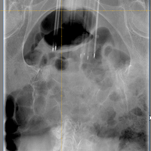
Elekta Studio enables fast, in-room imaging during treatment, allowing brachytherapy plans to be adapted to each patient’s daily anatomy with precision.
It provides detailed, artifact-free 2D and 3D images throughout the workflow—from applicator and needle navigation to treatment planning, verification, and delivery.
AI-enhanced imaging
Expand access to adaptive radiation therapy
High-definition AI-enhanced imaging will boost your confidence to deliver every radiotherapy dose with pinpoint accuracy.
Iris uses next-generation anatomy-specific AI scatter correction and Polyquant CT for radically improved CBCT image quality—without additional radiation dose.
*Iris® is a component of Elekta medical linear accelerators and has CE mark with limited global availability.
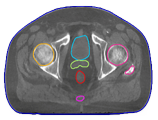
Elekta’s imaging upgrades provide cutting-edge solutions for today’s radiotherapy challenges:
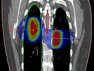
Revolutionary 4D imaging for motion management, enabling clinicians to track and adapt to respiratory motion with confidence.
Upgrade your CT-Linac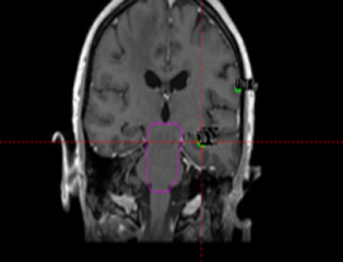
Advanced cranial SRS workflows, offering unparalleled precision and streamlined patient positioning.
Upgrade your Gamma Knife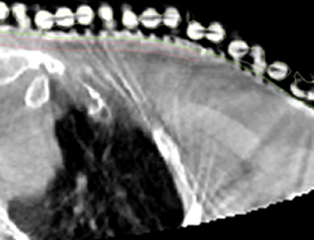
Seamless integration into workflows, enhancing accuracy and ease of planning.
Upgrade your Brachy workflow
AI-powered imaging delivering real-time insights, enhancing adaptive workflows, and improving treatment precision.
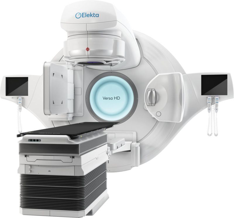
Upgrade your Versa HDElekta Care is your partner for success. Our people and technology keep you running reliably and efficiently while helping you to optimize outcomes and grow your practice.
We know integrating new imaging solutions into your workflow can be challenging, but you’re not alone. With practical application training and personalized peer-to-peer opportunities, we’ll help you unlock the full potential of your new solution from day one.
We’re here by your side to support you now and deliver a lifetime of high performance and progress.
About Elekta CareRequest more information from your local Elekta representative
Monaco, Agility and High Dose Rate Mode allow single fraction SABR treatment of a malignant lung neoplasm within a 20-minute treatment slot.
Read case study
“The CBCT-kV permitted us to identify patients for whom a positioning with bone-based 2D images only would have led to an unacceptable dose distribution. The easiness of implementing CBCT-kV imaging and its expected medical benefit should lead to a rapid diffusion of this technology.”
Pommier P, Gassa F, Lafay F, Claude L. Image guided radiotherapy with the Cone Beam CT kV (Elekta): experience of the Léon Bérard Centre. Cancer Radiother. 2009 Sep;13 (5):384-90. doi: 10.1016/j.canrad.2009.05.004. Epub 2009 Jul 28. PMID: 19640762. https://pubmed.ncbi.nlm.nih.gov/19640762/
“The requirements for 3D imaging became essential for as there was an increase in very sharp dose gradients adjacent to both targets and organs at risk, increasing the consequences of any geometric uncertainty, making daily treatment image verification (Image-guided radiation therapy—IGRT) an essential component of quality IMRT.”
Health.gov.au. MBS online - Item 15934: PET scan (Positron Emission Tomography) services. https://www9.health.gov.au/mbs/fullDisplay.cfm?type=item&q=15934&qt=ItemID
The perfect balance, without compromise
Push the boundaries of your stereotactic capabilities
Adapt at your rhythm
The only CT-Linac that uniquely evolves on your terms and offers everything from the everyday to the extraordinary
*Elekta Evo has CE mark with limited global availability.
Explore Evo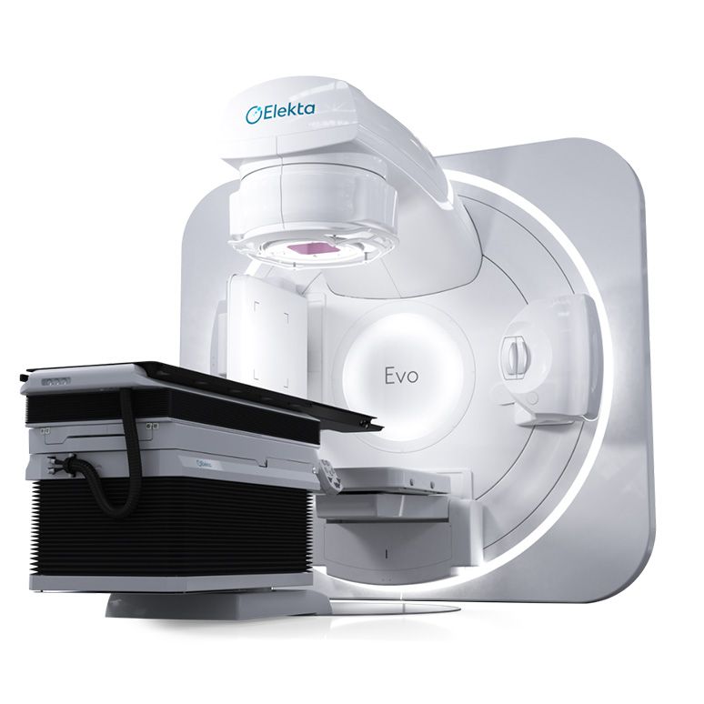
Protect the mind, protect the person
It allows high patient throughput with flexible workflows to ensure each patient’s treatment is personalized. Let’s talk about the advantages of upgrading to this exciting new platform.
Explore Esprit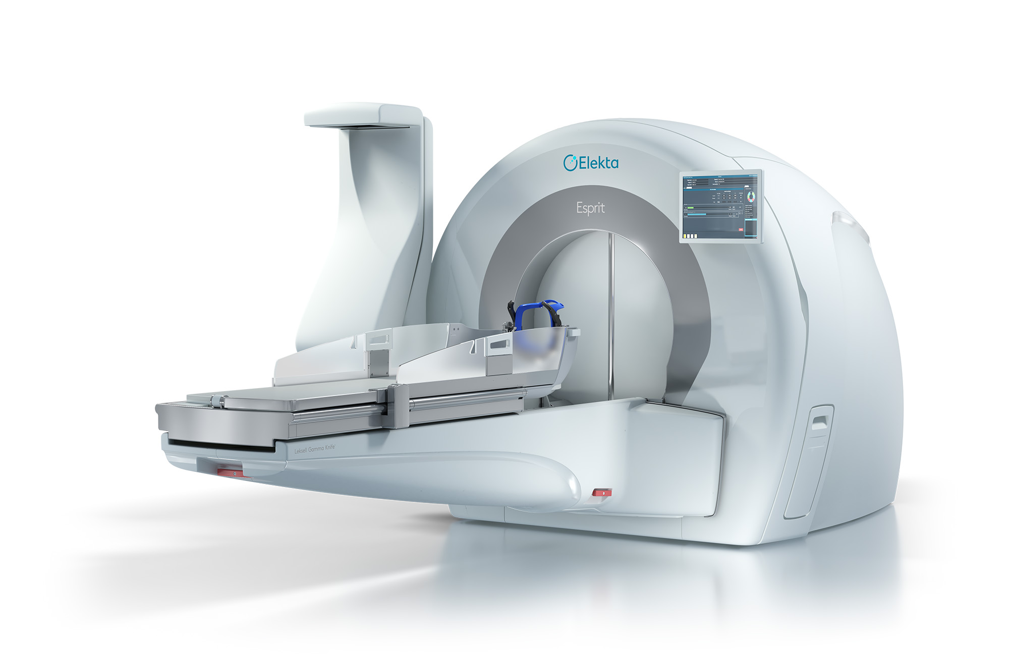
How MRI is propelling radiation therapy into a new era of clarity and insight.
Read white paper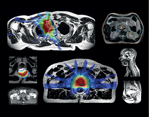

MR-guided radiotherapy can offer improved anatomical definition compared to on-board CBCT, while reducing radiation exposure.
Read case studySee the difference
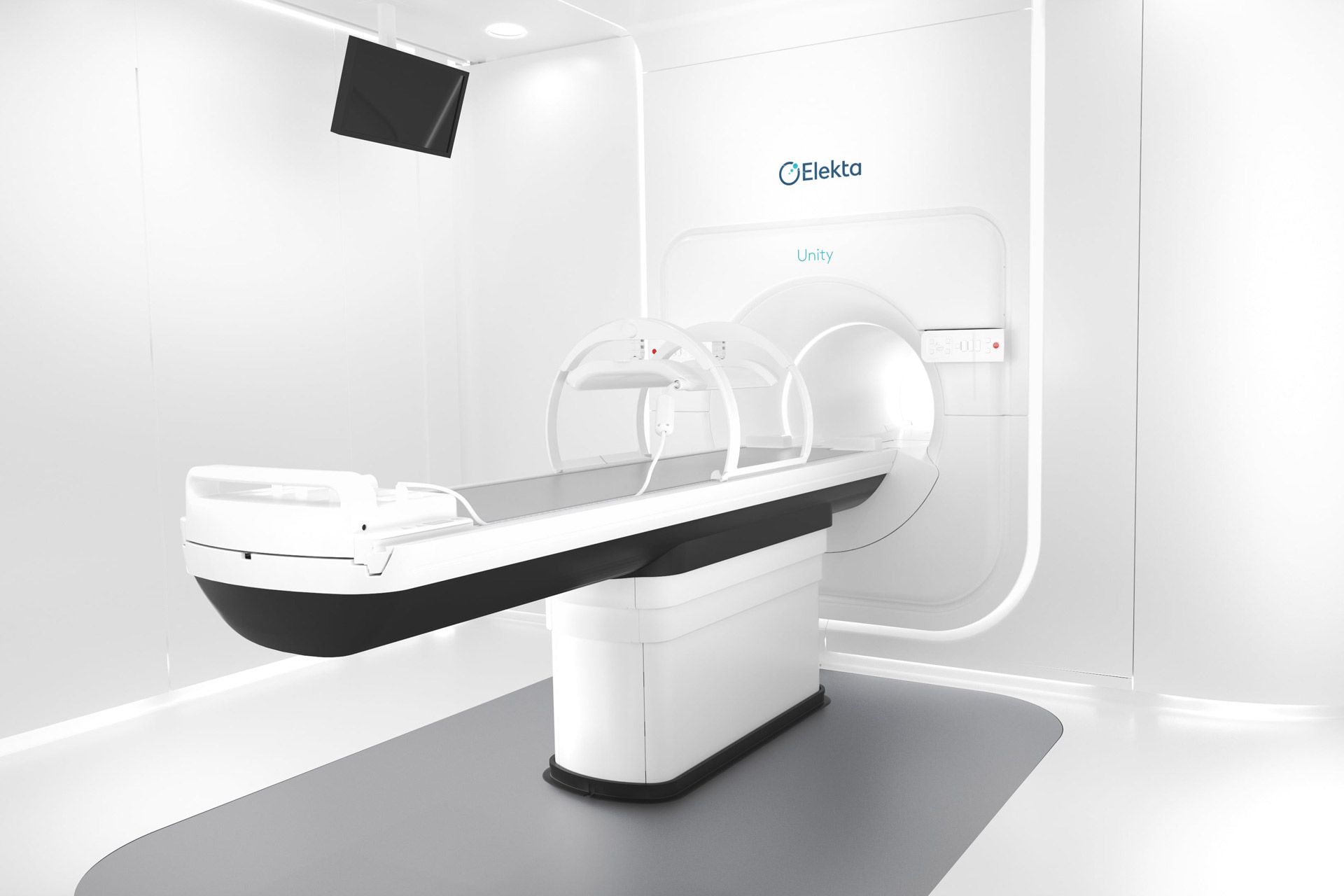

Discover how Comprehensive Motion Management (CMM) on Elekta Unity is enabling clinicians to confidently reduce margins.
Elekta offers a complete solution for contemporary interventional radiotherapy—bringing the entire brachytherapy workflow into one room.
Visit page View brochure