New Zealand clinicians deliver patient-friendly SABR lung in under two minutes

Treatment leverages Elekta’s Versa HD linac and Monaco Dynamic Conformal Arc Therapy

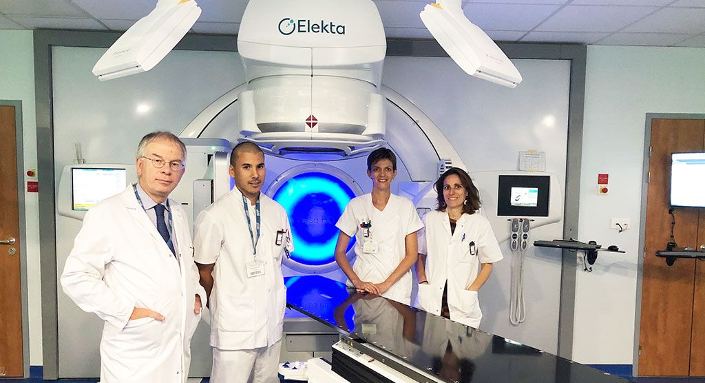
Lung cancer patients at Christchurch Hospital (Christchurch, New Zealand) have been benefitting from dramatically reduced radiotherapy treatment times since 2016, the year that the Canterbury District Health Board (CDHB) radiation oncology department invested in new technology. With its Versa HD™ linear accelerator and upgraded Monaco® (5.11) treatment planning system, clinicians have been delivering Monaco Dynamic Conformal Arc Therapy (DCAT) for its stereotactic ablative radiotherapy (SABR) lung patients, achieving a beam-on time of about 1.5 minutes.
It’s a major improvement over what the department had been using three years earlier when the SABR lung program started. SABR lung was performed using a 3D conformal radiotherapy (CRT) method with eight to nine noncoplanar beams. This method required patients to be on the treatment table for around one hour, including pre-treatment, mid-treatment and post-treatment imaging, which limited suitable patients to those who could tolerate this lengthy treatment time.
“Previously, SABR lung patients had to be able to tolerate 45 to 60 minutes on the treatment table,” says Andy Cousins, PhD, Radiation Oncology Physics Team Leader. “This restricted the treatment to fitter patients, who were few in number considering most of them were already unsuitable for surgery, and many required analgesics.
“SABR lung can now be delivered in a standard 15-minute time slot, including imaging.”

“With Monaco DCAT and Versa HD, treatment time has been reduced significantly,” Dr. Cousins continues. “SABR lung can now be delivered in a standard 15-minute time slot, including imaging. This means that today we can treat virtually all small NSCLC tumors using SABR and many patients are being referred for SABR lung as a first-choice treatment.”
SABR lung treatment at Christchurch Hospital delivers either 48 Gy in four fractions or 45 Gy in three fractions according to ESTRO Advisory Committee on Radiation Oncology Practice (ACROP) guidelines1. The margins currently used are 1.0 cm in the superior/inferior direction and 0.5cm everywhere else.
“Monaco DCAT provides the coverage without using VMAT,” he says. “The plans are easy and efficient to produce and are very robust for quality assurance (QA).”
Rapid treatment delivery
For treatment delivery, the patient is set up in the BodyFIX® vacuum bag. A Symmetry™ 4D CBCT is acquired to check the ITV and patient position, and automatic table adjustments are performed, if necessary, prior to starting treatment.
A 4D Symmetry scan is performed during treatment delivery. This eliminates the need for post-treatment CBCT imaging, which means that the patient can get off the table as soon as the treatment is finished.
“For SABR lung using DCAT and High Dose Rate mode, our typical beam-on time to deliver 15 Gy is just 1.5 minutes.”
“For SABR lung using DCAT and High Dose Rate mode, our typical beam-on time to deliver 15 Gy is just 1.5 minutes,” Dr. Cousins notes. “Imaging is acquired in just two to three minutes with an additional one to two minutes for analysis.
“The patient is on the table typically for 15 minutes, including set up and imaging, which is much easier for them to tolerate,” he continues. “We can book patients into a normal 15-minute treatment slot – rather than requiring three or four slots – so there is no interference with scheduling and we can treat more patients during the day.”

Advanced imaging techniques
Christchurch Hospital has been using Symmetry 4D CBCT for the management of respiratory motion since 2013. Symmetry provides anatomically correlated 4D image guidance at the time of treatment, providing volumetric visualization of moving tumors. No surrogates or external markers are required as Symmetry uses anatomical information contained within the projection data itself to automatically sort the projection images into the relevant phase “bins.”
The daily tumor position is then determined automatically by registering each phase to the reference image. By calculating displacement vectors for each registration, XVI understands the tumor trajectory and position of that specific day.
“When we started our SABR lung program, this seemed the ideal site to use Symmetry,” Dr. Cousins observes.

“Previously, we would have used 3D CBCT imaging,” he continues. “With Symmetry, we obtain a lot more information. Some small tumors that are not visible using 3D CBCT can be seen using Symmetry. In addition, we can see the tumor moving and we can check that the patient’s breathing pattern hasn’t changed since their planning CT scan. It also provides us with confidence about the accuracy of our treatments by accounting for baseline drift.”
Symmetry also provides valuable visual validation for the radiation therapists present at the time of treatment delivery.
“Symmetry gives us confidence that the movement captured in the 4D planning CT is accurate,” says Hayley Bennett, Radiation Therapist. “The vast majority of lesions move within the contoured ITV. It also fits easily into the workflow. We choose a clockwise or counter-clockwise Symmetry scan based on the side of the lesion. This makes the transition from the end of the Symmetry scan to the start of DCAT quick and easy.”
Recently, the department has also begun using 4D intrafraction imaging. This allows them to evaluate field coverage during treatment delivery and eliminates the need for a post-treatment CBCT scan, which reduces time on the table for the patient. Offline analysis of these images will also form part of a departmental study to explore the potential of margin reduction for SABR lung. They hope to be able to use this data to safely reduce the lung target margin to an isotropic 0.5 cm.
“We know where the tumor is located every day, for every fraction. We would not perform SABR lung treatments without it.”
“The combination of Symmetry and intrafraction imaging gives us confidence that we’re treating what we planned to treat,” Dr. Cousins says. “We know where the tumor is located every day, for every fraction. We would not perform SABR lung treatments without it.”
A better option for SABR lung patients
“Monaco DCAT has allowed the option of SABR lung treatment for patients who wouldn’t have been able to tolerate it previously due to the long treatment times,” Dr. Cousins says. “We are certainly treating more patients using this technique. Some of our lung surgeons are very keen for patients to have SABR lung, often as a first choice.”
“We are certainly treating more patients using this technique. Some of our lung surgeons are very keen for patients to have SABR lung, often as a first choice.”
“SABR lung is more effective and more convenient for patients than conventionally fractionated radiation treatment,” adds radiation oncologist Chris Harrington, MD. “Symmetry assures that tumor motion with breathing is accounted for in the treated volume, while DCAT and High Dose Rate mode provide a much quicker treatment time, which improves patient comfort and increases patient throughput. Few patients report more than minor side effects.”
“In the future, we’d like to expand our use of SABR for other soft tissue sites,” Dr. Cousins says. “Our experience with SABR lung and imaging will help enormously with that. We are hoping to start a trial on pancreas soon – our imaging solutions give us the confidence to do this as it could well involve a moving target.”
For more information on how Monaco DCAT SBRT compares to VMAT, click here.
- Guckenberger M, et al. ESTRO ACROP consensus guideline on implementation and practice of stereotactic body radiotherapy for peripherally located early stage non-small cell lung cancer. Radiother Oncol. 2017;124:10–17.


