MR-guided radiotherapy could open up the future to greater accuracy, personalized treatment and improved clinical research
By Kevin Brown, Distinguished Scientist, Elekta

Throughout the history of radiotherapy, scientists have worked hard to improve our ability to deliver the required dose of radiation to target while minimising side effects due to dose to the surrounding healthy organs. While major advances have been made our ability to see the target and healthy organs has been hampered by the performance of the imaging systems used at the time of treatment. MRI would be an obvious choice due to its superior ability to visualise soft tissues and lack of radiation exposure.
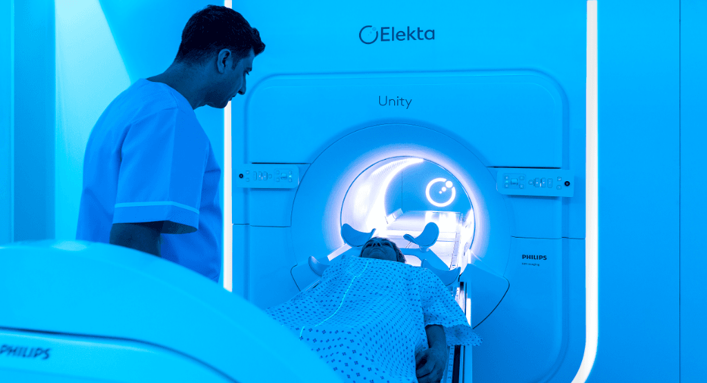
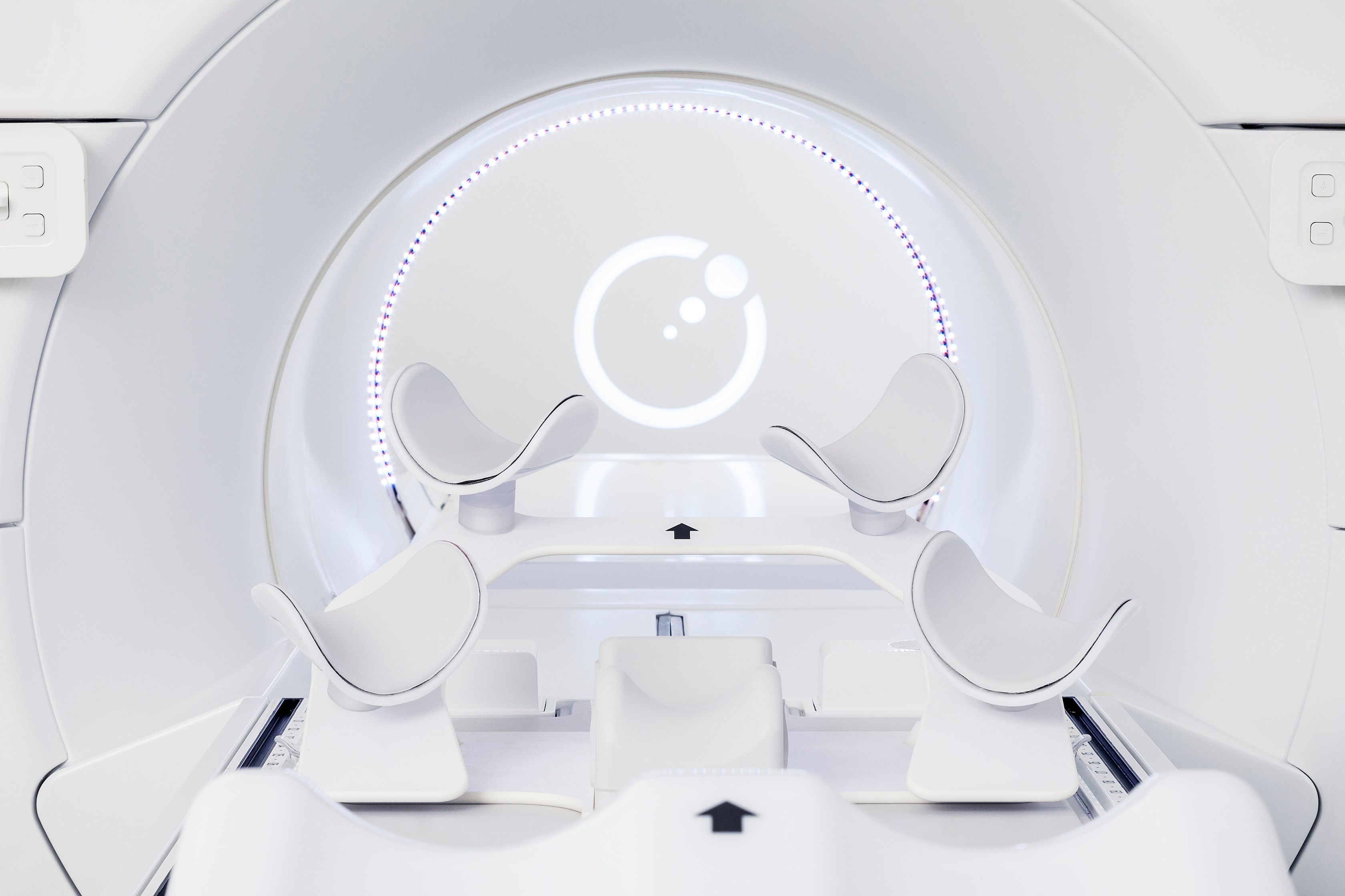
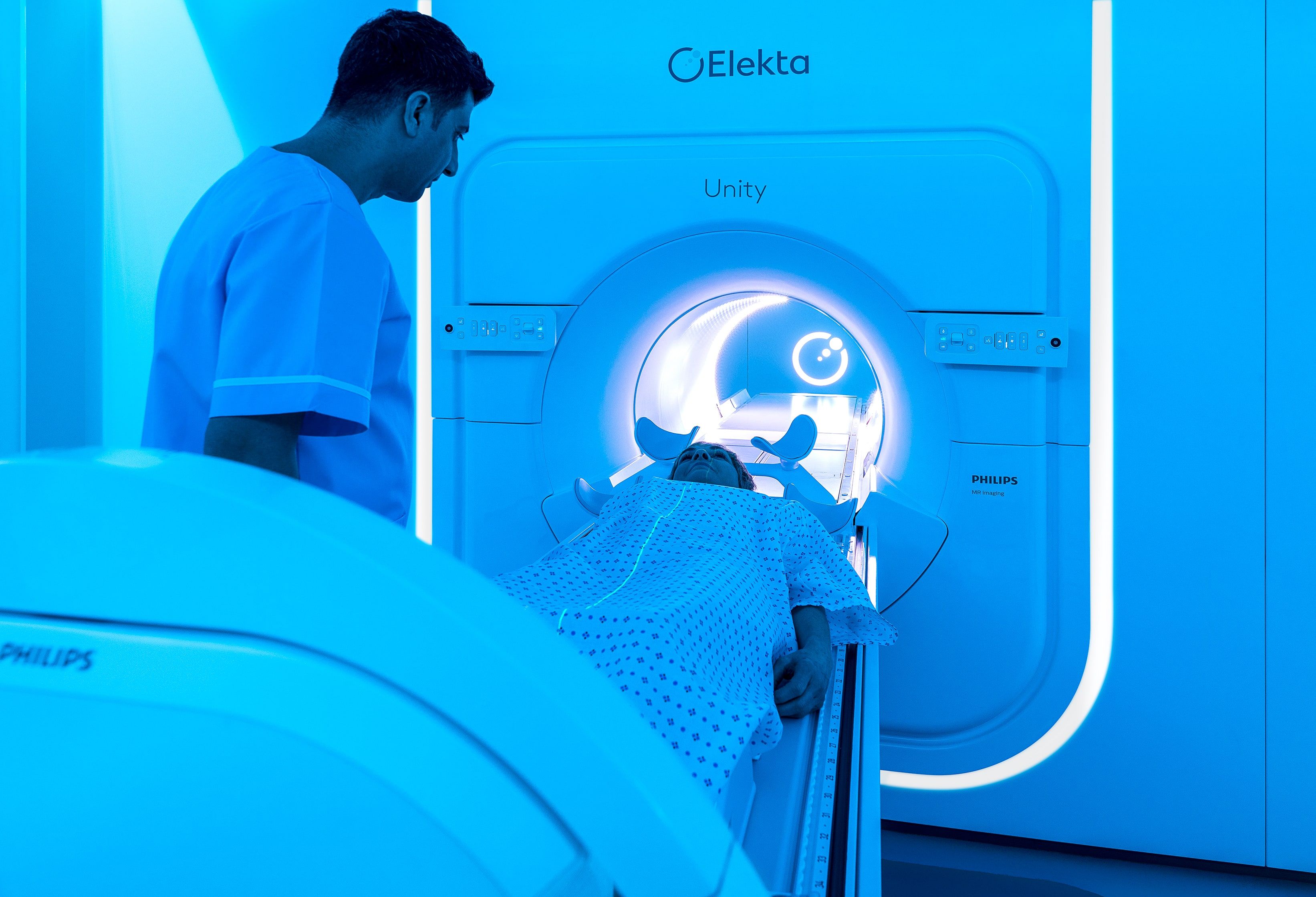
However, significant technical challenges are associated with the integration of an MRI, which is very sensitive to magnetic and RF interference, with a linac, which is also sensitive to magnetic interference. With Elekta Unity, we have worked with Philips to solve this challenge by creating a unique magnet which is designed to minimise the coupling between the two systems by using active shielding to create a low field annulus within which the linac gantry can rotate. This low field annulus not only allows the linac to operate in the presence of the MRI but also avoids any visible effect on the MRI images when the gantry is moving2. This means that the MRI can be used regardless of what the linac is doing. It also means that motion monitoring of the target does not need to be interrupted as the gantry moves from one beam to the next, also, that imaging biomarkers can be acquired simultaneously with the treatment being delivered.
The other key feature of Elekta Unity’s unique magnet is that it has been designed, as far as possible, to meet all the standard performance specifications that would be expected for diagnostic imaging. Our rationale for this is that significant advances are being made in mainstream MR technology and enabling radiotherapy to benefit from these is a significant opportunity. Elekta strongly believes that making these advanced capabilities available in the online setting through MR-guided therapy is going to be pivotal in the improvement of radiotherapy outcomes. Making Unity’s MRI very similar to mainstream diagnostic systems means that these MR innovations can be readily deployed onto Unity with minimal effort. Evidence of our success at doing this is the recent release of new MR techniques for managing motion described later in this article.
We have succeeded in creating the Elekta Unity MR-Linac which has exciting new capabilities which have previously been unavailable in radiotherapy.
“We have succeeded in creating the Elekta Unity MR-Linac which has exciting new capabilities which have previously been unavailable in radiotherapy.”
Integrating two systems creates high-quality tool for target motion management
Respiratory motion in patients is a challenge which we are tackling with Elekta Unity. Due to the integration of Philips mainstream MRI technology we have implemented three MR techniques to deal with respiratory motion, allowing clear and accurate images during free breathing, exhalation and breath holding:
- NAVIGATED – This approach automatically acquires a 3D image of the exhale respiration phase even though the patient is breathing freely. As the imaging is intermittent the image acquisition takes longer than a regular scan. However, the use of Philips C-SENSE technology can reduce this impact. This a fully automated process and does not require the use of a belt or a surface scanner to detect the breathing phase;
- 3D VANE – This imaging technique reduces motion artefacts in the images even though they are acquired while the patient is breathing freely. The resulting image is of the average target position and is likely to be appropriate for patients with small to moderate target motion;
- BREATHOLD – This is available in three different contrasts, T1, T2 and balanced, allowing the user to select the best target visibility for the clinical indication. A high-quality image can be provided within 18 seconds.
Because the MR has been so successfully isolated from the linac, image acquisition and target tracking can operate continuously throughout the treatment even when the gantry is moving. Also, as there is no radiation exposure associated with MR imaging there is no need to pause the imaging. Therefore, for the first time in the history of radiotherapy it is possible to continuously see the motion of the target in real time throughout the entire treatment delivery. This enables users to manage target motion while also being provided with highly accurate, real-time imaging to determine how much of the 3D target structure is outside the gating envelope at any one time.
Online plan adaptation can provide more personalised care, without moving the patient
Unity’s online plan adaptation also allows doses to be conformed to daily anatomy, supporting two different types of workflow – Adapt to Position and Adapt to Shape. Any of the daily changes in the position of the target can be compensated for by shifting the dose cloud to the new position rather than having to move the patient.
Avoiding moving the patient within the bore is beneficial in several ways. It allows the use of a wide comfort couch, avoids potential anxiety for the patients and means larger patients can be treated as it avoids the need for extra clearance around the patient to prevent collision.
In addition to improved patient experience, the primary rationale for this is looking to the future. In the future, we believe treatments will be fully adapted or dynamic and so simply providing a moving couch will add no value.
Elekta Unity supports precision treatment at every step
One of the concerns that people often express is about MR geometrical distortion. While it is possible to create MR images that have poor distortions it is also possible to manage these effects. Unity’s system design and approved MR sequences have been specifically designed to minimise distortions and a publication ref. demonstrated less than 1 mm distortion for a range of clinical targets that have been treated on Elekta Unity1 .
While creating Unity, we also devised specially designed tools and software that can facilitate specific quality assurance (QA) tasks. For example, measuring the alignment of the MR system and radiotherapy system is essential to ensure accurate dose delivery. Elekta provides specific phantom and software to perform this task. The phantom is imaged using the MRI and then using the integrated MV imager on the gantry allows the MV beam to be measured with respect to the phantom at all angles. The software then provides the calibration data to align the two systems to better than 1 mm. Because the two systems operate independently of one another there is no effect of the linac on the MRI and vice versa, therefore the MRI image quality and geometry QA is unaffected by the linac gantry.
Providing a platform for efficient clinical exploration
There is significant research suggesting that specific MR imaging biomarkers are good indicators of patient specific outcome, and this suggests that if those imaging biomarkers could be used to adapt the treatment then outcomes could be improved. However, research into these MR imaging biomarkers has been hindered by several factors:
- The cost and logistic challenges of acquiring the biomarker images
- The lack of biomarker standardisation
- The lack of coherent patient cohorts and follow up
Unity and the MR-Linac Consortium are addressing each of these barriers and we are optimistic that we are opening up a new and fruitful research domain. Unity’s diagnostic MRI can acquire many the imaging biomarkers of interest. Also, these imaging biomarkers can be acquired as part of a routine Unity treatment session eliminating the cost and logistic barriers – the patient is coming in for their treatment anyway.
The Imaging Biomarker group within the Consortium is developing standards for use across all Elekta Unity systems and benchmarking Unity against diagnostic MR systems. Finally, the patients that are being treated on the system are in coherent patient cohorts and are being followed up as part of the implementation of the Unity treatment technique and therefore this data will also be available for the biomarker research. We have implemented the MOMENTUM clinical registry which is becoming an unprecedented imaging biomarking repository, opening up a new future for clinical exploration in this exciting area.
Elekta Unity is opening up the future of efficiently assessing individual treatment response in an online setting and providing the ability to act on that response.
Learn more by watching my recent presentation.
Reference List
- Hasler, S. W., Bernchou, U., Bertelsen, A., van Veldhuizen, E., Schytte, T., Hansen, V. N., … Mahmood, F. (2020). Tumor-site specific geometric distortions in high field integrated magnetic resonance linear accelerator radiotherapy. Physics and Imaging in Radiation Oncology, 15, 100–104. https://doi.org/10.1016/j.phro.2020.07.007
- 10.1088/1361-6560/ab231a
