Elekta Studio ImagingRing brings improved control and precision to brachytherapy
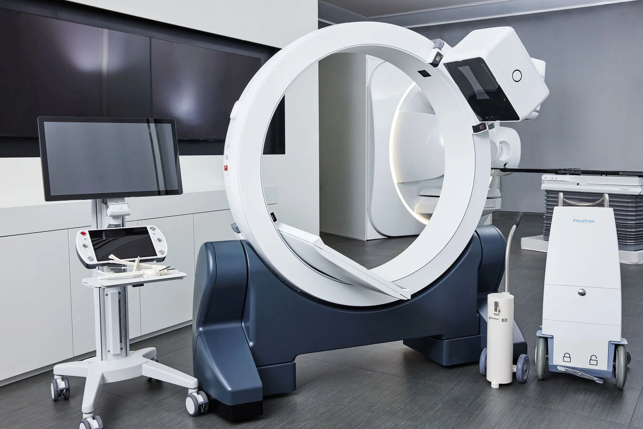
Physicists at Hong Kong’s Princess Margaret Hospital use solutions to streamline treatment workflow in image-guided brachytherapy
“Some types of cancer cannot be treated by external beam radiotherapy alone,” says Celia Chau, oncology physicist at Princess Margaret Hospital in Hong Kong. “But a combination of external beam radiotherapy and brachytherapy can really improve the outcome for the patient, and that’s why it’s so important.”
Getting the best from brachytherapy
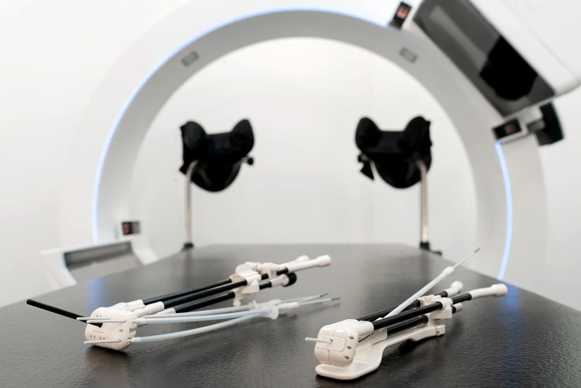
Having been using an Integrated Brachytherapy Unit (IBU) and 2D imaging for the last few years, the Princess Margaret Hospital has made the switch to Elekta Studio.
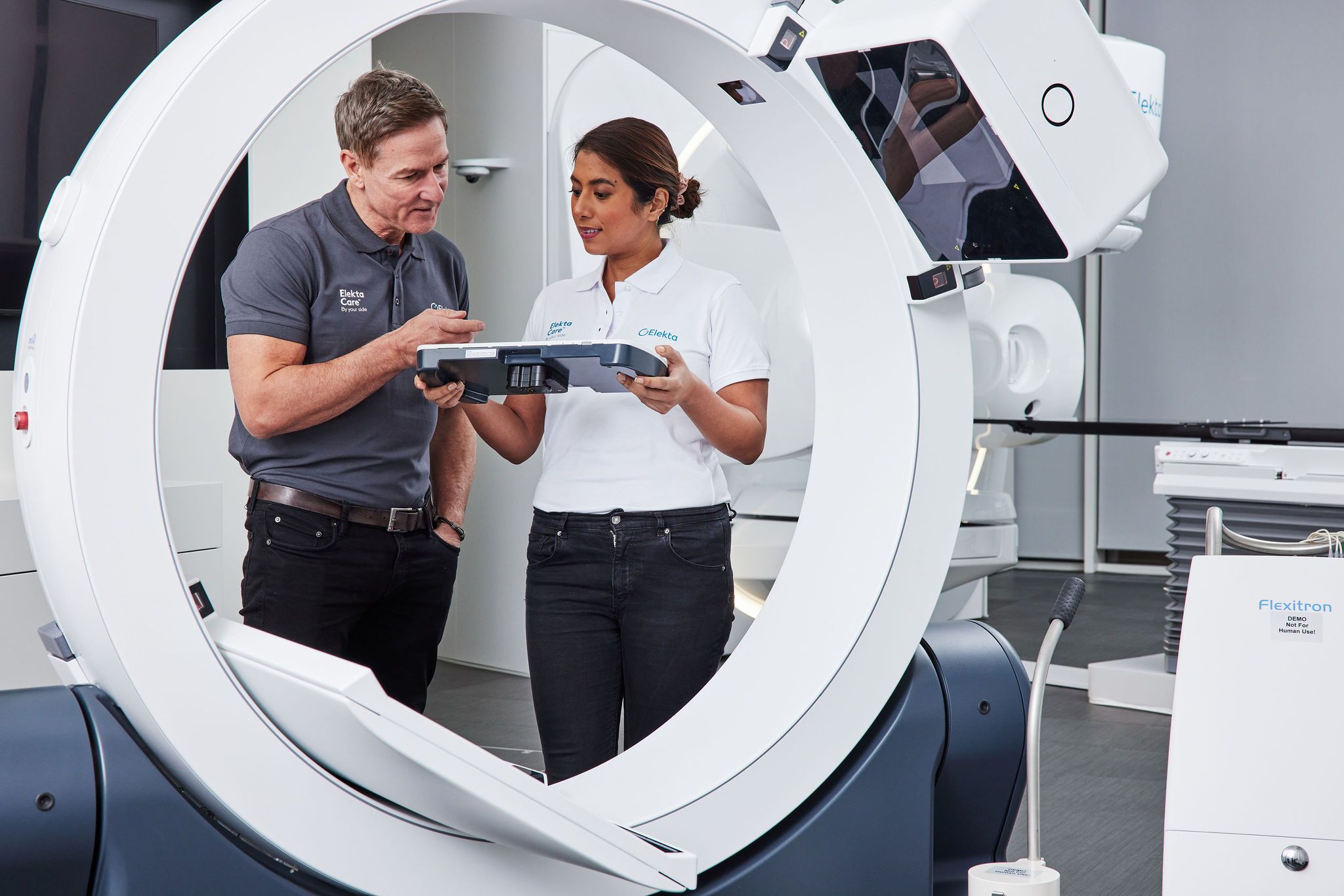
Elekta Studio is a suite of products designed for every step of the brachytherapy process, from applicators to imaging and treatment. One component of the solution, the ImagingRing, is a mobile CT scanner that enables clinicians to deliver all phases of the treatment from one room.

“If you have to move the patient to a separate imaging facility, you need to cover them and ask the nursing staff to transfer them back and forth between departments or buildings,” Ms. Chau explains. “But when everything is in one room, you don’t need that extra level of personnel. Every procedure is focused in the same place, and all the equipment is handy, so you have quick access to all needed tools.”
By reducing staff downtime and ensuring the appropriate staff and equipment are immediately accessible, caregivers can work more efficiently while maintaining a high patient care standard.
Streamlining workflows with 3D imaging
Exceptional quality imaging is vital to precision radiotherapy. The ImagingRing gives clinicians flexible imaging by virtue of a tilting gantry that allows them to scan patients in the treatment position.
“With an in-room 3D imaging option, the workflow has been much smoother than in our existing setting.”
“With an in-room 3D imaging option, the workflow has been much smoother than in our existing setting,” she says.

This is because a fully mobile CT scanner enables the treatment team to check and modify procedures in real time.
“After the doctor has inserted the applicator, they can immediately do the imaging and check whether it’s in the right location,” Ms. Chau observes. “And in between fractions, they can determine if the applicator has moved. If it has, they can immediately adjust the applicator or use the planning system to reacquire the image and then adapt the plan.”
Delivering efficient quality assurance
Minimal movement and strong visibility reduce uncertainty. This means high doses of radiation can be used with confidence, knowing that the tumor is optimally targeted, while still protecting nearby organs at risk.
“As a physicist, I always have to consider the applicator displacement inside the patient, because high dose rate brachytherapy is not without risks,” she notes. “If the applicator moves slightly — even just one or two millimeters – that could significantly change the tumor dose and dose coverage.”
Improving patient outcomes
Patients are more likely to feel comfortable if they can stay in one room for all aspects of their treatment, rather than being moved from place to place. And with a spacious scanner and advanced imaging capability, it is easier to acquire the right images in the correct position and at the right time. This all helps the procedure progress smoothly, reducing the patient’s anxiety and making the experience as stress-free as possible.
“Based on the existing market for a mobile imaging system, the image quality is superior.”
“Based on the existing market for a mobile imaging system, the image quality is superior,” Ms. Chau says. “Because it’s protocol-based, everything is based on the individual patient, accounting for things like their age or weight. Therefore, the operator doesn’t have to use trial and error to determine the exposure setting for the best image. It’s quite an amazing thing.”
With the right equipment, clinicians can deliver efficient and streamlined patient experiences. And they can create effective, personalized plans, which reduce treatment time, aid recovery, and minimize the impact on quality of life.
Supporting brachytherapy
Elekta Studio is just one of the ways Elekta is advancing brachytherapy practices. The company is also raising awareness with professionals, whether they are just getting started or extending their skills and knowledge.
The BrachyAcademy offers observational visits, workshops, on-site consultancy training, and a wealth of information from a variety of publications. It brings medical and clinical expertise into care teams, drawing on more than 35 years of experience and innovation.
When caregivers can design treatments that emphasize patient comfort, they can deliver a different kind of brachytherapy. Elekta Studio gives clinicians instant images at any time throughout the patient’s treatment, informing every stage from planning to treatment delivery. And it can all be done in the same place, making the experience easier for staff and less stressful for patients.
“You get everything in one room, which means greater certainty with treatment.”
“You get everything in one room, which means greater certainty with treatment,” Ms. Chau concludes. “And that gives me more confidence to do my job.”
Learn more about 3D image-guided brachytherapy with Elekta Studio.
*Elekta Studio is comprised of multiple medical devices, some of which may not yet be available in all markets. Elekta is the authorized Exclusive Distributor of medPhoton in Brachytherapy. ImagingRing is product manufactured by medPhoton GmbH. ImagingRing may not be available in all markets.
LWBBX230106
