Consortium demonstrates phantom’s value in SRS end-to-end accuracy

In Elekta-sponsored webinar, presenters describe how phantom enables more accurate stereotactic radiosurgery
Verifying the end-to-end accuracy of 3D dose delivery for stereotactic radiosurgery (SRS) is critical due to the technique’s small margin of error, according to Emmanouil Zoros, PhD, medical physicist at RTsafe (Athens, Greece), during a webinar hosted by Physics World.

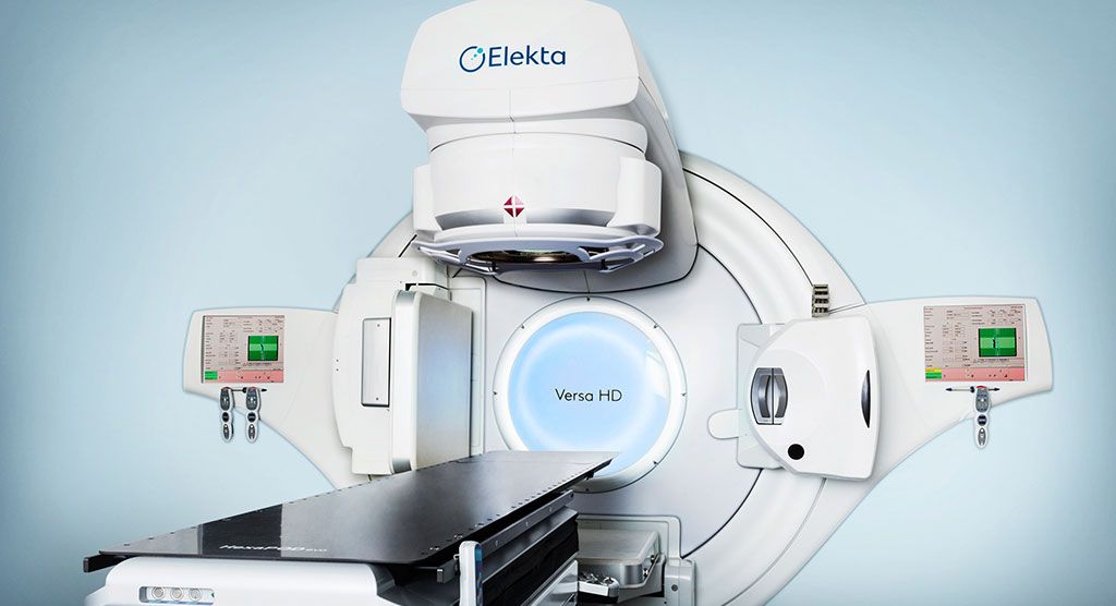
Dr. Zoros’s talk was followed by a presentation given by Dr. Daniel L. Saenz (University of Texas Health Science Center), who provided results of a six-site consortium that clinically validated an innovative RTsafe phantom prototype. The centers used the Elekta’s Versa HD™ system, HexaPOD™ evo-RT patient positioning and Monaco® treatment planning in their evaluation.
“Quality assurance in SRS demands a high level of accuracy and precision, focusing on dosimetry and geometry,” Dr. Zoros said. “It’s crucial to verify the accuracy of whole SRS delivery chain, from imaging to dose delivery, and that can be accomplished with an appropriate phantom. RTsafe’s PseudoPatient™ Prime human anatomy phantom fulfills key requirements for compatibility with various imaging modalities and patient immobilization, contrast quality in MR and CT, physical dimensions and other important aspects. This enables it to provide an end-to-end evaluation of advanced radiotherapy verification focusing on SRS.”

The PseudoPatient Prime approach combines highly accurate, medical grade 3D printing technology and the latest dosimetry methodologies, he explained. The phantom uses CT datasets of real patients, 3D construction of cranial bone anatomy with density corresponding to bone equivalent materials, and the option of water or gel as tissue equivalent materials.
Consortium tests RTsafe phantom
Following Dr. Zoros’ presentation, Daniel L. Saenz, PhD, Assistant Professor of Medical Physics, University of Texas Health Science Center (San Antonio, Texas, USA), discussed the results of a six-site international consortium, which took on the challenge of validating a prototype of RTsafe’s PseudoPatient phantom in the clinic. Each of the centers used an Elekta Versa HD™ system for high definition dynamic radiosurgery (HDRS), all of which were equipped with the Agility™ MLC and HexaPOD evo-RT robotic patient positioning system. Plans were created with Monaco.
“The RTsafe phantom enables us to make a dosimetric 3D measurement that is as clinically realistic as possible,” Dr. Saenz observed. “The primary advantage is that we’re not simply recalculating in a phantom geometry, which is what physicists do all the time. With the PseudoPatient phantom, we are able to measure the dose with a geometry that is identical to that of the patient. That means no recalculations on a different phantom, we’re simply treating the plan exactly as if it were the patient, and we can compare the actual clinical dose distribution rather than some recalculated dose distribution as it would lay in the phantom.”
With the PseudoPatient phantom, we are able to measure the dose with a geometry that is identical to that of the patient.
Each site received a phantom and a CT dataset that included a set of structures to plan on; the CT used in the study was a patient-derived scan.

“The patient had multiple intracranial targets ranging in size from six millimeters to 25 millimeters, representing a typical range of targets that we would expect to treat,” he said. “Each target was prescribed a dose of 8 Gy, and then allowing a peaking hot spot dose within the target, which was meant to stay below 12 Gy. In addition to the brain mets, we added a somewhat larger QA target that we treated with a homogenous dose of 8 Gy.”
The centers did a CT scan of the phantom to fuse it with the planning CT and ensure that they achieved the described agreement. They treated the phantoms (ion chamber, gel and film phantoms) as if there were the patient.
“They were set up on the Versa HD system, aligned to the planning isocenter using XVI CBCT and then positioned using the HexaPOD positioning system,” Dr. Saenz noted. “Across all institutions, we found agreement within 1.7 percent with an average agreement of 1.2 percent. Most importantly in terms of accuracy, we found that within a distance of four centimeters from the isocenter, we found the highest gamma passing rates and we also found spatial agreements that were constrained within one millimeter. In other words, for all targets within four centimeters of the isocenter, this would correspond to a sphere with a diameter of eight centimeters. Beyond four centimeters from the isocenter we saw a modest increase from one millimeter.”
Overall, Dr. Saenz observed, the investigators found agreement between the measurement and calculation that was typical of conventional SRS end-to-end testing. Compared with single-isocenter, single-target SRS, there was agreement within four centimeters. Point measurement results were within two percent, and film measurement gamma analysis was dependent on film placement relative to the targets. 3D gel dosimetry is the preferred tool for single isocenter, multi-metastasis, including the ability to look at gamma analysis, DVH and dose statistics.
He concluded by noting:
- Isocenter is a point, we are showing SRS-type accuracy within an eight-centimeter sphere around the isocenter point, making single isocenter, multi-mets treatment feasible on Elekta HDRS linacs.
- Measuring end-to-end accuracy is critical to understanding how well a system performs stereotactic treatments and the results here easily demonstrate the accuracy necessary for confident treatments on Elekta HDRS linacs.


